A knee X-ray is a quick imaging test that uses a small amount of radiation to look at the bones and joint inside your knee.
🦴 What a knee X-ray shows
A knee X-ray mainly shows bone structures, including:
- Thigh bone (femur)
- Shin bone (tibia)
- Smaller bone (fibula)
- Kneecap (patella)
- Joint space between bones
It does NOT clearly show soft tissues like ligaments, cartilage, or muscles.
📸 Why doctors order it
A knee X-ray is commonly used for:
- 🦴 Fractures (broken bones)
- 🧓 Arthritis (joint wear and tear)
- 🦵 Knee pain or swelling
- 🧱 Bone deformities
- 🏃 Injury after falls or sports trauma
🧪 Types of knee X-ray views
Doctors may take different angles:
- AP view (front view)
- Lateral view (side view)
- Oblique view (angled view)
- Weight-bearing X-ray (checks joint space under pressure)
⚙️ What a normal knee X-ray looks like
- Smooth bone edges
- Even space between bones
- No cracks or fractures
- No abnormal bone growth
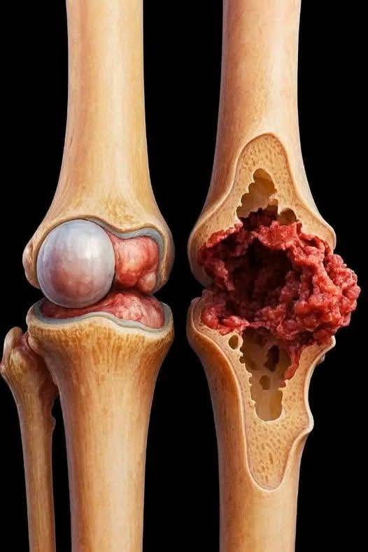
⚠️ What abnormal findings may show
- Narrow joint space → possible arthritis
- Fracture lines → broken bone
- Bone spurs → osteoarthritis
- Swelling signs around joint (indirect)
🩻 Is it painful or risky?
- ❌ Not painful
- ⚡ Very low radiation exposure
- ⏱️ Takes only a few minutes
- 👕 No special preparation needed
✔️ Bottom line
A knee X-ray is a simple and fast test used mainly to check bones and joint damage, especially after injury or in arthritis cases.
If you want, I can also show you what arthritis vs normal knee looks like on X-ray or explain when MRI is better than X-ray 👍
