An X-ray of the knee is a medical imaging test used to look at the bones and joint structure of the knee.
🦴 Knee X-ray
Knee X-ray
📸 What it shows
A knee X-ray mainly shows bones, not soft tissues. It can help doctors see:
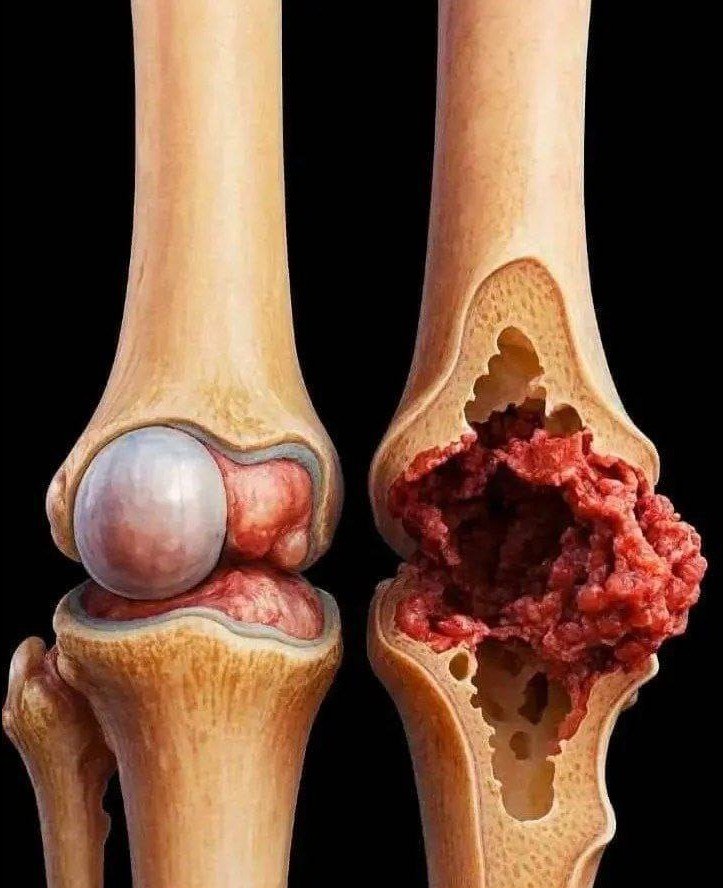
- 🦴 Bone fractures (breaks)
- 🧱 Arthritis (joint space narrowing)
- 🦵 Bone alignment problems
- 🪨 Bone spurs (extra bone growth)
- ⚖️ Joint wear and degeneration
🏥 When it is done
Doctors may order a knee X-ray for:
- Knee pain or swelling
- Injury or fall
- Difficulty walking
- Suspected arthritis
- Chronic stiffness
⚙️ How it works
- You place your knee on an X-ray table
- A small amount of radiation passes through the body
- Dense structures (bones) appear white on the image
⚠️ Important facts
- Quick and painless
- Very low radiation exposure
- Does not show ligaments, tendons, or cartilage clearly (MRI is better for that)
🧠 Simple summary
A knee X-ray is a fast and simple scan that shows bone problems in the knee joint, mainly used for fractures and arthritis.
If you want, I can also show:
🧠 Difference between X-ray vs MRI for knee pain
🦵 Common knee problems seen on X-rays
📊 Normal vs arthritis knee X-ray explanation in simple images
