Here is what an X-ray of the knee joint typically looks like and what it is used for:
🦴 What a knee X-ray shows
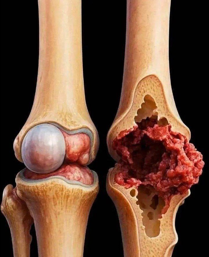
A knee X-ray mainly shows the bone structure of the joint, including:
- Femur (thigh bone)
- Tibia (shin bone)
- Patella (kneecap)
It helps doctors see alignment and detect bone-related problems.
🧠 Common reasons for a knee X-ray
🦴 1. Arthritis
- Narrowing of joint space
- Bone spurs
- Common in older adults
⚡ 2. Injury or fracture
- Cracks or breaks in bone
- After falls or accidents
⚖️ 3. Joint alignment problems
- Misalignment or wear-and-tear changes
💧 4. Swelling assessment (indirect)
- Shows changes around the joint area
🏥 When doctors recommend it
- Knee pain or stiffness
- Injury or trauma
- Difficulty walking
- Suspected arthritis
⚠️ Important note
- X-rays show bones clearly, but not soft tissues like ligaments or cartilage
- For soft tissue injuries, MRI is usually needed
✔️ Bottom line
A knee X-ray is a basic imaging test used to check bones, joint alignment, fractures, and arthritis in the knee.
If you want, I can show you normal vs arthritic knee X-rays or explain what your specific knee pain might mean 👍
