Here is what an X-ray of the knee joint looks like and what it shows.
🦴 What a knee X-ray is
A knee X-ray is a medical imaging test that uses low-dose radiation to create pictures of the bones in the knee joint, mainly:
- Femur (thigh bone)
- Tibia (shin bone)
- Patella (kneecap)
It is commonly used to check bone alignment and joint damage.
🧠 What doctors look for
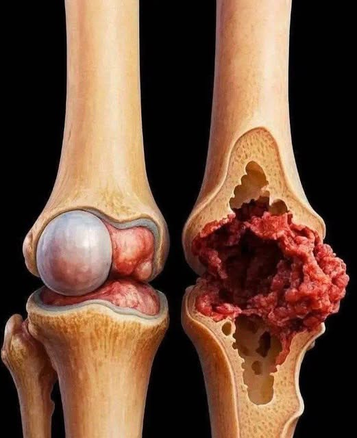
🟡 1. Arthritis
- Joint space narrowing
- Bone spurs
- Common in older adults
🦴 2. Fractures
- Breaks or cracks in bones
- Often after injury or falls
⚖️ 3. Joint alignment problems
- Misalignment of bones
- Wear and tear changes
💧 4. Fluid or swelling (indirect signs)
- Soft tissue changes around the joint
🏥 When it is recommended
- Knee pain or swelling
- Injury or trauma
- Difficulty walking
- Suspected arthritis
⚠️ Important note
- X-rays show bones clearly, but not soft tissues like ligaments or cartilage
- For those, doctors may use MRI instead
✔️ Bottom line
A knee X-ray is a simple and fast scan that helps doctors see bone problems, fractures, and signs of arthritis in the knee joint.
If you want, I can also show normal vs arthritic knee X-rays or explain knee pain causes based on X-ray findings 👍
